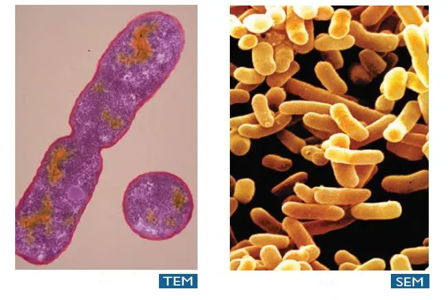
Difference between Scanning Electron Microscopy (SEM) and Transmission Electron Microscopy (TEM)

The advent of Electron Microscopy in 1932 open the door to visualize small subcellular structure and viruses which were beyond the scope of Light microscopy which can’t resolve objects separated by less than 0.2μm.
Facts: Ernst Ruska has got Nobel prize in Physics in 1986 for his fundamental work in electron options, and for the design of the first electron microscope, other half of the prize was jointly given to Gerd Binnig and Heinrich Rohrer “for their design of the scanning tunneling microscope”.

Electron microscope uses the beam of electrons which travel through a vacuum and electromagnets are used to focus the beam. Though electron microscopes are expensive and requires much more space for installation of the equipment, the electron micrographs it generates justify the capital investments. Greater details of minute biological structures generated through EM is aiding in the advancement of research breakthroughs.
Five basic steps involved in all Electron Microscopes are:
- A stream of high voltage electrons (usually 5-100 KeV) is formed by the electron source (usually a heated tungsten or field emission filament) and accelerated in a vacuum toward the specimen using a positive electrical potential.
- This stream is confined and focused using metal apertures and magnetic lenses into a thin, focused, monochromatic beam.
- This beam is focused onto the sample using electromagnetic lens.
- Interactions occur inside the irradiated sample, affecting the electron beam.
- These interactions and effects are detected and transformed into an image.
There are several types of electron microscopes, classified according to their final use. Transmission electron microscope and scanning electron microscope are two most common types of electron microscope.

The major differences between SEM and TEM are as follows:
| Properties | Scanning Electron Microscopy (SEM) | Transmission Electron Microscopy (TEM) |
| Light Source | SEM is based on scattered electrons, i.e. electrons emitted from the surface of a specimen. It is the EM analog of a stereo light microscope. | Electrons are used as “light source”. TEM is based on transmitted electrons and operates on the same basic principles as the light microscope. |
| Purpose | SEM provides detailed images of the surfaces of cells. SEM focuses on the sample’s surface and its composition, so SEM shows only the morphology of samples. | Transmission electron microscope is used to view thin specimens (tissue sections, molecules, etc). TEM can show many characteristics of the sample, such as internal composition, morphology, crystallization etc. |
| Sample Preparation | Sample is coated with a thin layer of a heavy metal such as gold or palladium. | The sample in TEM has to be cut thinner (70-90 nm) because electrons cannot penetrate very far into materials. |
| Resolution | SEM can resolve objects as close as 20 nm. | TEM has much higher resolution than SEM. It can resolve objects as close as 1 nm i.e. down to near atomic levels. |
| Magnification | The magnifying power of SEM is up to 50,000X. | The magnifying power of TEM is up to 2 million times. |
| Processing of sample (s) | SEM allows for large amount of sample to be analyzed at a time | With TEM only small amount of sample can be analysed at a time. |
| Image formation |
Secondary or backscattered electrons arising from interaction of electron beam and metal coated specimen are collected and the resulting image is displayed on a computer screen.
| Transmitted electrons hit a fluorescent screen giving rise to a “shadow image” of the specimen with its different parts displayed in varied darkness according to their density. The image can be studied directly by the operator or photographed with a camera. |
| 3D picture | SEM provides a 3-dimensional image | TEM provides a 2-dimensional picture. |
| Current Applications | To study the topography and atomic composition of specimens, process control and also, for example, the surface distribution of immuno-labels | To image the interior of cells (in thin sections), the structure of protein molecules (contrasted by metal shadowing), the organization of molecules in viruses and cytoskeletal filaments (prepared by the negative staining technique), and the arrangement of protein molecules in cell membranes (by freeze-fracture). |
References:For more information please read/ visit:
- Microbiology: Principles and Explorations, 9th Edition; Jacquelyn G. Black, Laura J. Black
- https://www.umassmed.edu/cemf/whatisem/
- https://www.va.gov/DIAGNOSTICEM/What_Is_Electron_Microscopy_and_How_Does_It_Work.asp
- https://www.nobelprize.org/educational/physics/microscopes/tem/