Quantitative Buffy Coat (QBC) Test: Principle, Method and Analysis

Two methods, thick and thin blood smear microscopy are regarded as the “gold standard” for the diagnosis of malaria. Quantitative Buffy Coat is another direct and rapid test for diagnosis of malaria. It is based on acridine orange staining of centrifuged peripheral blood samples in a microhematocrit tube (QBC) and examination under UV light source (fluorescence microscopy).
The acridine orange stains all nucleic acid containing cells and the associated fluorescence is observable under blue-violet light through a microscope.
According to the manufacturer, QBC Malaria Test is 5.5 to 7% more sensitive than Giemsa thick films. It can detect as little as 1 parasite per μL of blood and establish diagnosis earlier than thick film in 47% of low parasitemia (<10 parasites per μL) cases.
QBC is established as an effective tool for diagnosing blood parasites that cause malaria, filariasis and visceral leishmaniasis.
Principle:
Acridine orange binds deoxyribonucleic acids and ribonucleic acids. The malaria parasite binds acridine orange in the nucleus and the cytoplasm and emits green and red fluorescence when excited by blue light (at 460 nm) allowing the detection and examination of parasite morphology by fluorescent microscopy. The nuclei of the parasites emit yellowish green fluorescence whereas the cytoplasm exhibits bright red fluorescence. RBCs are not stained by the dye, hence remain inconspicuous under fluorescent light (dark background) while the brightly fluorescent parasites are easily seen. The outlines of stained parasites are well preserved and the general morphology is similar to that in specimens stained by the Giemsa stain.
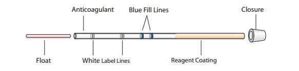
Sample collection: Blood sample can be collected in either capillary finger-prick or phlebotomy in a ethylenediamine tetraacetate (EDTA) containing vials.
About the QBC tube:
The QBC glass capillary tube (Becton Dickinson) is 75 mm in length and 1.677 mm in diameter. The tubes are internally coated with EDTA and heparin at the fill end and with acridine orange stain and potassium oxalate at the other end.
Procedure:
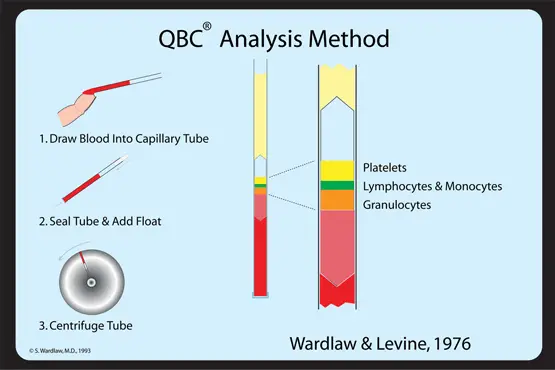
- Draw samples of blood ( 55 µl) in to the QBC tube by capillary action.
- Rotate the tubes for 10 seconds to dissolve the contained residues in the blood.

- Insert a close fitting cylindrical insert or plastic float { having a specific gravity (1.055) i.e midway between that of plasma (1.028) and red blood cells (1.090)} inside a acridine orange-coated capillary tube.
- Centrifuge the tubes at 12,000 g for 5 minutes.
After gentrification blood components and malaria parasites separate based on density, and concentrate in distinct layers.
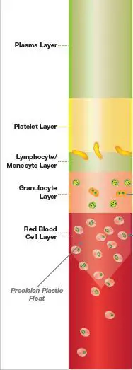
Note: The float by virtue of its density settles on top of the centrifuged packed red cells. It occupies 90% cross-sectional area of the tube which aids in the expansion of the centrifugally seperated cell layers. It is surrounded by three discernible and now measurable layers of the buffy coat. - Insert the centrifuged QBC Malaria test into the Paraviewer. Position the tube so the closure end extends over the depressed area of the holder.
- The area surrounding the float just beneath the buffy coat was examined under oil immersion. Individual cells within this layer were easily seen by microscopy; the malaria parasites staining green (DNA) and orange (RNA) under blue-violet light.
- The entire circumference of the tube was examined systematically while moving away from the buffy coat through the erythrocyte layer.
- Each tube was examined until parasites were detected or for a maximum of 5 minutes.
Note: You can download this QBC malaria test user guide to get step by step guideline.
Results/Analysis
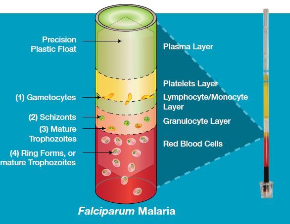
If a sample contains P. falciparummalaria parasites:
- Crescent shaped gametocytes (1) will appear near the interface of the lymphocyte/monocyte and platelet layers.
- A small number of (2) schizonts and (3) mature trophozoites may appear in the granulocyte layer.
- Ring-shaped (4) immature trophozoites will appear throughout the red blood cell layer, with a concentration near the interface with the granulocyte layer.
Other parasites species, including P. vivax, will also concentrate
during centrifugation, but exhibit different characteristics
during centrifugation, but exhibit different characteristics
Acridine Orange Staining: Principle, Procedure, Results and Applications
Acridine orange is a dye that intercalates or binds with the nucleic acid ( either DNA or RNA) present in organisms and fluoresce to emit various colors that help in differentiation of cellular organells. This binding is the result the electrostatic interactions of acridine molecule between the nucleic acid base pairs. Acridine Orange (AO), due to its metachromatic properties, is commonly used in fluorescence microscopy and flow cytometry analysis of cellular physiology and cell cycle status, including the fluorescent microscopic examination of microorganisms.
Principle : Acridine orange is a cell-permeable,nucleic acid selective dye that emits green fluorescence when bound to dsDNA(at 520 ) and red fluorescence when bound to ssDNA or RNA(at 650 nm).Since it is a cationic dye,it also enter acidic compartments such as lysosomes which in low pH conditions, will emit orange light.
Staining procedure:
Staining procedures vary according to its use
- For staining clinical specimen with acridine orange at low pH (Acridine orange Acid Stain)
- Requirements: Acridine orange,Glacial Acetic acid, Distilled water
- Preparation of reagent: 50 mg acridine orange is dissolved in 10 ml of distilled water to preparea stock solution and stored in the refrigerator.1 ml of Acridine orange stock solution and 0.5 ml of glacial acetic acid is added to 50 ml of distilled water to prepare a working solution.
- Staining procedure:
- Prepare a smear in a clean grease free slide and allow it to air dry.
- The slide is then fixed with methanol and dried again.
- It is then put in trough with acridine orange staining working solution (i.e 0.01 per cent).
- After 2 minutes of staining, the slides are washed gently with water and dried and then examined in a fluorescent microscope.Observance: Bacteria stain orange against a green to yellow background of human cells and debris.
- For staining cells for analysis by flow cytometry.
Requirements: 0.1M Citric Acid (dissolve 1.921g per 100ml distilled water) ,0.2M Dibasic Sodium Phosphate (dissolve 2.839g per 100ml distilled water) ,Triton X-100 (Baker),0.5M EDTA, Sodium chloride(NaCl), Acridine Orange (Powder) and Sucrose. - Preparation of reagents:
- Stock Buffer I :20mM Citrate-Phosphate, pH 3.0, 0.1mM EDTA, 0.2M Sucrose, 0.1% Triton X-100
(To 125ml distilled water add 40µl 0.5M EDTA, 26.48ml 0.1M Citric Acid, 6.85ml 0.2M Dibasic Sodium Phosphate, 13.69g Sucrose, 0.2ml Triton X-100 .QS to 200ml and 0.2µ filter. Store at 4oC)
Stock Buffer II :10mM Citrate-Phosphate, pH 3.8, 0.1M NaCl - (To 150 ml distilled water add 9.92ml 0.1M Citric Acid, 5.46ml 0.2M Dibasic Sodium Phosphate, 1.7g NaCl. QS to 200ml and 0.2m filter. Store at 4oC)
- Stock Buffer I :20mM Citrate-Phosphate, pH 3.0, 0.1mM EDTA, 0.2M Sucrose, 0.1% Triton X-100
Staining Procedure :
- Make a 2mg/ml solution of Acridine orange in distilled water and dilute to 1:100 in Buffer II
- Aliquot cells: 105- 106 in 100µl PBS or media .
- Add Buffer I (0.5ml) at room temp, agitate to suspend .
- Add Buffer II + AO (0.5ml) at room temp, agitate to suspend.
- Run on flow cytometer. Excitation 488 nm; dot plot of green fluorescence at 530nm versus red fluorescence >600 nm).
Observance:
Green fluorescence when bound to dsDNA and red fluorescence when bound to ssDNA or RNA.
Applications:
- For analyzing mitochondria and lysosomal content by flow cytometry.
- For visual detection of nucleic acids on agarose and polyacrylamide gels.
- For enumerating the microbial load in a sample since acridine orange binds with the nucleic acid of both living and dead bacteria.
- For identifying engulfed apoptotic cells, because it will fluoresce upon engulfment.
- For differential staining of human cells and prokaryotic cell with a fluorescence microscope. Human cells are stained black to faint green in which Bright orange organisms are easily detected .
Researches showed that
Acridine orange staining is a sensitive, rapid and reliable method for detecting bacteria in blood cultures early during incubation and can be substituted for blind subcultures.
Acridine orange is better than Gram stain in cases with low amounts of organisms.