To identify and differentiate bacteria based on chemical composition of cell wall
One of the best differential staining method followed in microbiology to identify bacteria is Gram staining. The method was introduced by a Danish Physician Hans Christian Gram in 1884. This method differentiates and classifies bacteria based on chemical composition of the cell wall. It is also used to find size, shape and arrangement of bacteria.
Principle of Gram Staining: The bacterial cell wall is composed of peptidoglycon and lipopolysaaccharides
clinically important bacteria can be detected/visualized using Gram staining method the only exceptions being those organisms;
That exists almost exclusively within host cells i.e. Intracellular bacteria (e.g., Chlamydia)
Those that lack a cell wall (e.g., Mycoplasma)
Those of insufficient dimensions to be resolved by light microscopy (e.g., Spirochetes)
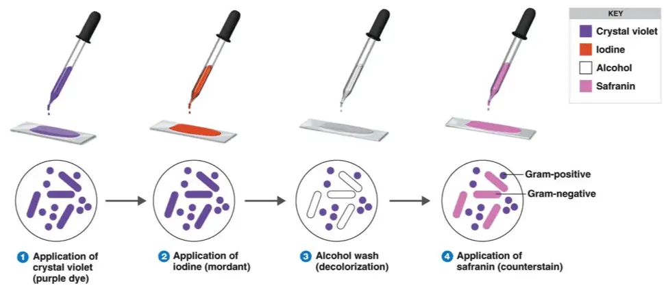
Classic Gram staining techniques involves following steps:
Fixation of clinical materials to the surface of the microscope slide either by heating or by using methanol. (# Methanol fixation preserves the morphology of host cells, as well as bacteria, and is especially useful for examining bloody specimen material).
An easy way to remember the steps of the Gram stain
Application of the primary stain (crystal violet). Crystal violet stains all cells blue/purple
Application of mordant: The iodine solution (mordant) is added to form a crystal violet iodine (CV-I) complex; all cells continue to appear blue.
Decolorization step: The decolorization step distinguishes gram-positive from gram-negative cells.
The organic solvent such as acetone or ethanol, extracts the blue dye complex from the lipid-rich, thin walledgram negative bacteria to a greater degree than from the lipid poor, thick walled, gram-positive bacteria. The gram negative bacteria appear colorless and gram positive bacteria remain blue.
Application of counter stain (safranin): The red dye safranin stains the decolorized gram-negative cells red/pink; the gram-positive bacteria remain blue.
Principle of Gram Stain
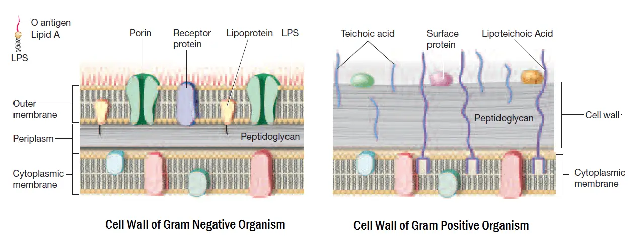
Image 2: Cell wall of Gram Positive and Gram Negative Bacteria
The differences in cell wall composition of Gram positive and Gram negative bacteria accounts for the Gram staining differences. Gram positive cell wall contain thick layer of peptidoglycan with numerous teichoic acid cross linking which resists the decolorization.
In aqueous solutions crystal violet dissociates into CV+ and Cl – ions that penetrate through the wall and membrane of both Gram-positive and Gram-negative cells. The CV+ interacts with negatively charged components of bacterial cells, staining the cells purple.
When added, iodine (I- or I3-) interacts with CV+ to form large crystal violet iodine (CV-I) complexes within the cytoplasm and outer layers of the cell.
The decolorizing agent, (ethanol or an ethanol and acetone solution), interacts with the lipids of the membranes of both gram-positive and gram negative bacteria.
The outer membrane of the Gram-negative cell (lipopolysaccharide layer) is lost from the cell, leaving the peptidoglycan layer exposed. Gram-negative cells have thin layers of peptidoglycan, one to three layers deep with a slightly different structure than the peptidoglycan of gram-positive cells. With ethanol treatment, gram-negative cell walls become leaky and allow the large CV-I complexes to be washed from the cell.
The highly cross-linked and multi-layeredpeptidoglycan of the gram-positive cell is dehydrated by the addition of ethanol. The multi-layered nature of the peptidoglycan along with the dehydration from the ethanol treatment traps the large CV-I complexes within the cell.
After decolorization, the gram-positive cell remains purple in color, whereas the gram-negative cell loses the purple color and is only revealed when the counterstain, the positively charged dye safranin, is added.
Smear Preparation
Fix material on slide with methanol or heat. If slide is heat fixed, allow it to cool to the touch before applying stain.
Image 3: Procedure of Gram Staining; note the color change after each steps
Gram Staining Procedure/Protocol:
Flood air-dried, heat-fixed smear of cells for 1 minute with crystal violet staining reagent. Please note that the quality of the smear (too heavy or too light cell concentration) will affect the Gram Stain results.
Wash slide in a gentle and indirect stream of tap water for 2 seconds.
Flood slide with the mordant: Gram’s iodine. Wait 1 minute.
Wash slide in a gentle and indirect stream of tap water for 2 seconds.
Flood slide with decolorizing agent (Acetone-alcohol decolorizer). Wait 10-15 seconds or add drop by drop to slide until decolorizing agent running from the slide runs clear .
Flood slide with counterstain, safranin. Wait 30 seconds to 1 minute.
Wash slide in a gentile and indirect stream of tap water until no color appears in the effluent and then blot dry with absorbent paper.
Observe the results of the staining procedure under oil immersion (100x) using a Bright field microscope.
If you are struggling to remember the staining reagents used in this procedure and their order you can remember this sentence “Come In And Stain” i.e. the order is Crystal violet, Iodine, Alcohol/Acetone and the final one is Safranin.
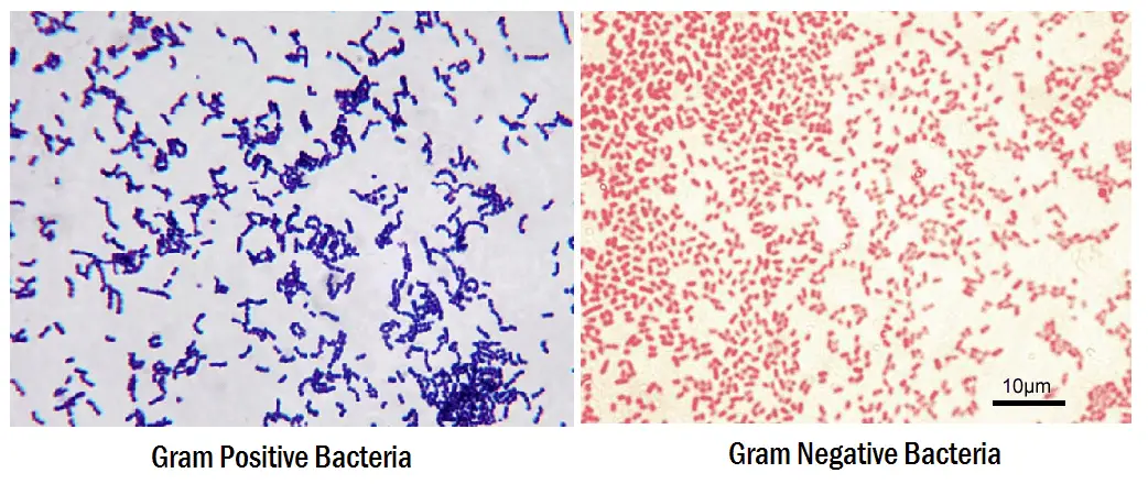
Results:
Gram-negative bacteria will stain pink/red and
Gram-positive bacteria will stain blue/purple.
Reporting Gram smears
The report should include the following information:
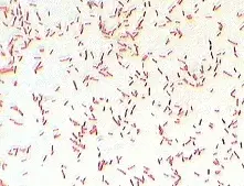
Staphylococcus in Gram Stain
Numbers of bacteria present, whether many, moderate, few, or scanty
Gram reaction of the bacteria, whether Gram positive or Gram negative
Morphology of the bacteria, whether cocci, diplococci, streptococci, rods, or coccobacilli. Also, whether the organisms are intracellular.
Presence and number of pus cells
Presence of yeast cells and epithelial cells.
Example
An urethral smear report might read: ‘Moderate numbers Gram negative intracellular diplococci and many pus cells.’
Quality Control: Always check new batches of stain and reagents for correct staining reactions using a smear containing known Gram positive and Gram negative organisms.
The structure of the organism’s cell wall determines whether the organism is gram psitive or negative. When stained with a primary stain and fixed by a mordant, some bacteria are able to retain the primary stain by resisting declorization while others get decolorized by a decolorizer. Those bacteria which retain the primary stain are called Gram positive and those bacteria which gets decolorized and then get counterstained are called Gram negative. Crystal violet (CV) dissociates into CV+ and Cl– ions in aqueous solutions. These ions penetrate through the cell wall and cell membrane of both Gram-positive and Gram-negative cells. The CV+ ion interacts with negatively charged components of bacterial cells and stains the cells purple.
Iodine (I), used as mordant interacts with CV+ and forms large complexes of crystal violet and iodine (CV–I) within the inner and outer layers of the cell.
When a decolorizer such as alcohol or acetone is added, it interacts with the lipids of the cell membrane. Since Gram negative organism have thin peptidoglycan layer(1-2 layers) and have additional lipopolysaccharide layer which gets dissolved due to the addition of alcohol, so gram negative organism fails to retain the complex and gets decolorized as the complex is washed away.
In contrast, a Gram-positive cell becomes dehydrated from an ethanol treatment. This closes the pores in the cell wall and prevents the stain from exiting the cell. The large CV–I complexes become trapped within the Gram-positive cell also due to the thick and multilayered (40 layers) nature of its peptidoglycan.
After decolorization, the Gram-positive cell remains purple and the Gram-negative cell loses its purple color. Counterstain, which is usually positively-charged safranin or basic fuchsin, is applied last to give decolorized Gram-negative bacteria a pink or red color.
REQUIREMENTS AND PREPARATION OF REAGENTS
Primary Stain : Crystal violet
Solution A :
Crystal violet = 2 gm
Ethyl alcohol= 20 ml
Solution B :
Ammonium oxalate = 0.8 gm
Distilled water = 80 ml
Mix solution A and B. Keep for 24 hours and filter. Store in an amber colored bottle.
Mordant : Gram’s Iodine
Iodine = 1 gm
Potassium iodide = 2 gm
Distilled water = to 100 ml
Mix and Store in an amber colored bottle.
Decolorizer : 95% Ethanol or 1:1 acetone with ethanol
Acetone = 50 ml
Ethanol (95%) = 50ml
Counterstain: safranin
Safranin O = 0.34 gm
Absolute alcohol = 10ml
Distilled water = 90ml
Mix, filter and store in ambered colored bottle.
PROCEDURE OF GRAM STAINING
Smear preparation :
Take a grease free dry slide.
Sterilize the inoculating loop on a flame of a Bunsen burner.
Transfer a loopful of culture (or the specimen) by sterile loop and make a smear at the center. Smear should not be very thin or very thick.
Allow the smeat to dry in the air.
Fix the dry smear by passing the slide 3-4 times through the flame quickly with the smear side facing up.
Gram Staining :
Place the slides on the staining rods.
Cover the smear with crystal violet stain and leave for 1 minute.
Wash carefully under running tap water.
Flood the smear with Gram’s iodine solution and leave for 1 minute.
Drain off the iodine Wash the slide for the again in a gentle stream of tap water.
Flood the slide with the decolorizing agent then wait for 20-30 seconds. This can also be done by adding a drop by drop to the slide until the decolorizing agent running from the slides runs clear.
Gently wash the slide under running tap water and drain completely.
Counterstain with safranin for and and wait for about 30 seconds to 1 minute.
Wash slide in a gentile and indirect stream of tap water until no color appears in the effluent and then blot dry with absorbent paper.
Observe under microscope.
INTERPRETATION OF GRAM STAINING
The staining results of gram stain are as follows :
Gram staining is a simple, differential staining method which helps to group bacteria according to their Gram character (Gram positive or Gram negative). It is one of the initial steps carried out in pathological laboratories for the identification of etiological agent and hence is one of the most important staining techniques in medical microbiology. The Gram reaction depends on the growth phase of the organism, young and growing bacteria gives most consistent reaction.
Principle - Gram staining depends on the structure and composition of the cell wall of bacteria. Gram positive bacteria have thicker peptidoglycan layer than gram negative bacteria. First, primary stain, crystal violet stains all the cells purple. Addition of iodine (mordant) forms crystal violet iodine complex within the cell wall. The complex formed is larger than crystal violet so it cannot be easily washed out from the intact peptidoglycan layer. Application of alcohol (decolorizer) decolorizes the stain. Since gram negative organism have thin peptidoglycan layer and have additional lipopolysaccharide layer which gets dissolved due to the addition of alcohol, so gram negative organism fails to retain the complex and gets decolorized . On other hand gram positive continues to retain the complex and remain purple. To observe the decolorized cells secondary stains like Basic fuchsin or Safranin is added which stains the gram negative organisms pink.
Procedure
1. Prepare the smear and heat fix it.
2. Add crystal violet (primary stain) - 1minute.
3. Wash and add Gram’s iodine (mordant) - 1 min.
4. Drain off iodine, add alcohol (decolorizer) -15 seconds
5. Wash and add Basic fuchsin or Safranin (secondary stain)- 1 min.
6. Wash, dry and observe under oil immersion lens.
Observation
Purple cells - Gram positive
Pink cells – Gram negative
Gram staining also helps in studying the morphological features and cellular arrangements of the organisms.
Gram staining method, the most important procedure in Microbiology, was developed by Danish physician Hans Christian Gram in 1884. Gram staining is still the cornerstone of bacterial identification and taxonomic division.
This differential staining procedure separates most bacteria into two groups on the basis of cell wall composition:
Gram positive bacteria (thick layer of peptidoglycan-90% of cell wall)- stains purple
Gram negative bacteria (thin layer of peptidoglycan-10% of cell wall and high lipid content) –stains red/pink
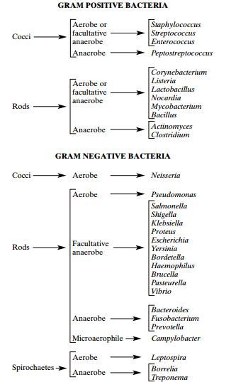
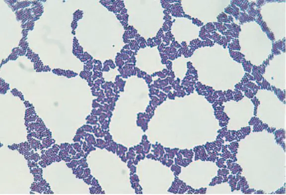
Image 1: Basic classification of Medically Important Bacteria
Nearly all clinically important bacteria can be detected/visualized using Gram staining method the only exceptions being those organisms;
That exists almost exclusively within host cells i.e. Intracellular bacteria (e.g., Chlamydia)
Those that lack a cell wall (e.g., Mycoplasma)
Those of insufficient dimensions to be resolved by light microscopy (e.g., Spirochetes)
Classic Gram staining techniques involves following steps:
Fixation of clinical materials to the surface of the microscope slide either by heating or by using methanol. (# Methanol fixation preserves the morphology of host cells, as well as bacteria, and is especially useful for examining bloody specimen material).
An easy way to remember the steps of the Gram stain
Application of the primary stain (crystal violet). Crystal violet stains all cells blue/purple
Application of mordant: The iodine solution (mordant) is added to form a crystal violet iodine (CV-I) complex; all cells continue to appear blue.
Decolorization step: The decolorization step distinguishes gram-positive from gram-negative cells. The organic solvent such as acetone or ethanol, extracts the blue dye complex from the lipid-rich, thin walled gram negative bacteria to a greater degree than from the lipid poor, thick walled, gram-positive bacteria. The gram negative bacteria appear colorless and gram positive bacteria remain blue.
Application of counter stain (safranin): The red dye safranin stains the decolorized gram-negative cells red/pink; the gram-positive bacteria remain blue.
Principle of Gram Stain
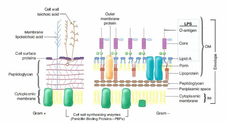
Image 2: Cell wall of Gram Positive and Gram Negative Bacteria
The differences in cell wall composition of Gram positive and Gram negative bacteria accounts for the Gram staining differences. Gram positive cell wall contain thick layer of peptidoglycan with numerous teichoic acid cross linking which resists the decolorization.
In aqueous solutions crystal violet dissociates into CV+ and Cl – ions that penetrate through the wall and membrane of both Gram-positive and Gram-negative cells. The CV+ interacts with negatively charged components of bacterial cells, staining the cells purple.
When added, iodine (I- or I3-) interacts with CV+ to form large crystal violet iodine (CV-I) complexes within the cytoplasm and outer layers of the cell.
The decolorizing agent, (ethanol or an ethanol and acetone solution), interacts with the lipids of the membranes of both gram-positive and gram negative bacteria.
The outer membrane of the Gram-negative cell (lipopolysaccharide layer) is lost from the cell, leaving the peptidoglycan layer exposed. Gram-negative cells have thin layers of peptidoglycan, one to three layers deep with a slightly different structure than the peptidoglycan of gram-positive cells. With ethanol treatment, gram-negative cell walls become leaky and allow the large CV-I complexes to be washed from the cell.
The highly cross-linked and multi-layeredpeptidoglycan of the gram-positive cell is dehydrated by the addition of ethanol. The multi-layered nature of the peptidoglycan along with the dehydration from the ethanol treatment traps the large CV-I complexes within the cell.
After decolorization, the gram-positive cell remains purple in color, whereas the gram-negative cell loses the purple color and is only revealed when the counterstain, the positively charged dye safranin, is added.
Smear Preparation
Fix material on slide with methanol or heat. If slide is heat fixed, allow it to cool to the touch before applying stain.
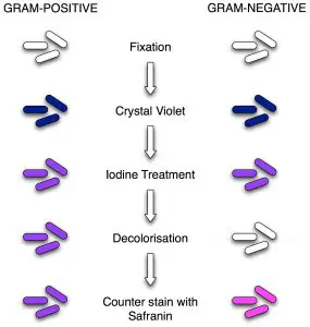
Image 3: Procedure of Gram Staining; note the color change after each steps
Gram Staining Procedure/Protocol:
Flood air-dried, heat-fixed smear of cells for 1 minute with crystal violet staining reagent. Please note that the quality of the smear (too heavy or too light cell concentration) will affect the Gram Stain results.
Wash slide in a gentle and indirect stream of tap water for 2 seconds.
Flood slide with the mordant: Gram’s iodine. Wait 1 minute.
Wash slide in a gentle and indirect stream of tap water for 2 seconds.
Flood slide with decolorizing agent (Acetone-alcohol decolorizer). Wait 10-15 seconds or add drop by drop to slide until decolorizing agent running from the slide runs clear .
Flood slide with counterstain, safranin. Wait 30 seconds to 1 minute.
Wash slide in a gentile and indirect stream of tap water until no color appears in the effluent and then blot dry with absorbent paper.
Observe the results of the staining procedure under oil immersion (100x) using a Bright field microscope.
If you are struggling to remember the staining reagents used in this procedure and their order you can remember this sentence “Come In And Stain” i.e. the order is Crystal violet, Iodine, Alcohol/Acetone and the final one is Safranin.
Results:
Gram-negative bacteria will stain pink/red and
Gram-positive bacteria will stain blue/purple.
Reporting Gram smears
The report should include the following information:
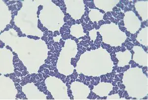
Staphylococcus in Gram Stain
Numbers of bacteria present, whether many, moderate, few, or scanty
Gram reaction of the bacteria, whether Gram positive or Gram negative
Morphology of the bacteria, whether cocci, diplococci, streptococci, rods, or coccobacilli. Also, whether the organisms are intracellular.
Presence and number of pus cells
Presence of yeast cells and epithelial cells.
Example
An urethral smear report might read: ‘Moderate numbers Gram negative intracellular diplococci and many pus cells.’
Quality Control: Always check new batches of stain and reagents for correct staining reactions using a smear containing known Gram positive and Gram negative organisms.
Difference between Gram positive and Gram Negative bacterial cell wall
The cell wall of Gram negative bacteria is more complex than those of Gram positive bacteria. Gram negative bacteria contain an extra layer of cells called outer membrane or LPS layer which surrounds the thin peptidoglycan layer. LPS layer is absent in Gram positive bacteria.
Some of the main differences between Gram positive bacteria and Gram negative bacteria are:
Staphylococcus in Gram StainGram Stain Reaction of E.Coli
Properties
Gram Positive Bacteria
Gram Negative Bacteria
Thickness of cellwall
Thicker than Gram negative bacteria. around 20 to 25 nm
Generally thinner, 11 to 15 nm
Gram reaction
Gram positive bacteria stain a deep blue color (violet/purple) in Gram staining technique
Gram negative bacteria stain pink to red color in Gram staining technique.
Thick (multilayered) peptidoglycan layer is present in Gram positive bacteria. It accounts 50% or more of the dry weight of the wall of some Gram positive bacteria.
Thin (single-layered). Around 10% weight of the cellwall of Gram negative bacteria.
Teichoic acids
Cell wall of gram positive bacteria bacteria contains teichoic acids.
Absent
Periplasmic space
Periplasmic space is absent in Gram positive bacteria
There are two periplasmic space in Gram negative bacteria; one between the murein and inner cell membrane and the other between the murein and outer cell membrane.
Primarily endotoxins, LPS layer has a endotoxic property.
Lipid content
Low
High around 11 to 22% of dry weight of the cell wall (because of lipid rich LPS layer).
Action of Lysozyme
Cell wall of Gram positive bacteria is easily destroyed by the action of lysozyme. After digestion of Peptidoglycan layer, Gram positive bacteria become protoplast.
Gram negative bacteria are refractory to lysozyme, because large protein molecule cannot penetrate the LPS layer. After digestion of Peptidoglycan layer, Gram negative bacteria become spheroplasts.
Spheroplasts: Gram negative bacteria with intact cytoplasmic membrane of the protoplast plus the outer membrane (LPS layer) of the cell wall , after peptidoglycan layer is destroyed by lysozyme or its synthesis inhibited by antibiotics.
Protoplasts: Cells whose walls have been completely remove and are incapable of normal growth and division.
I hope you all are well aware about the Gram staining, its protocol (procedure), principles and interpretation. In this blog post, I am writing some other important aspects of Gram Stain which is less discussed or less memorized by most of the students, but which is useful and worth knowing.
We all know that Gram stain is the most important staining technique for identifying bacteria using light microscopy but Gram staining techniques also have some limitations. We know how to perform gram stain but we may not know the procedure we applied is adequate or not.
You may fail to recover organism in Gram Stain from clinical specimen but same clinical specimen yields organisms when cultured. So lets find why this happens.
Limitation of Gram Stain: Mycobacteria stain weakly with gram stain and bacteria such as Mycoplasma, Rickettsiae, Chlamydiae do not take up the dyes used in Gram stain or are too small to be seen with light microscopy.
Sensitivity of Gram Staining Technique: To be visible on a slide, organisms that stain by the Gram method must be present in concentrations of about 10^4 to 10^5 organisms per milliliter of uncentrifuged fluid.
Adequacy of Gram Staining method: After performing gram stain, Microbiologist/Technician should first determine whether the Gram stain is adequate. In an appropriately stained specimen, the nuclei of neutrophils are red. If the nuclei are blue, the decolorization is insufficient.
Variations in Gram Reaction
Gram positive bacteria may lose their ability to retain crystal violet and stain Gram negatively for the following reasons:
Cell wall damage of bacteria due to antibiotic therapy or excessive heat fixation of the smear.
Over- decolorization of the smear
Use of an Iodine solution which is too old, i.e. yellow instead of brown in color (always store in a brown glass or other light opaque container).
Smear has been prepared from an old culture.
When smear is too thick, Gram negative bacteria may not be fully decolorized during decolorization steps and appear as Gram positive bacteria.
Importance of Gram Stain in Anaerobic Bacteriology
Gram stain is an important rapid tool for anaerobic bacteriology. It reveals the types and relative numbers of microorganisms and host cells present. Gram stain also serves as a quality control measure for the adequacy of anaerobic techniques.
Is there any modification of the Gram staining technique for the anaerobes?
Yes. We use the same standard gram stain procedure and reagents but the safranin counterstain is left on for 3 to 5 minutes. Alternatively, 0.5% aqueous basic fuchsin can be used as the counterstain.
What does positive gram stain and negative culture indicates?
Positive grams stain with negative culture report gives information regarding adequacy sample collection, transport and also of culture methods used. This situation may come in the following mentioned conditions:
Poor transport methods
Excessive exposure to air during sample processing
Inadequate types of media or old media, or
Failure of the anaerobic system used (jar, pouch, and chamber) to achieve an anaerobic atmosphere.
That microorganisms have been killed by antimicrobial therapy