The negative stain is particularly useful for determining cell size and arrangement. It can also be used to stain cells that are too delicate to be heat-fixed. Nigrosin is an acidic stain.
Negative staining is an established method, often used in diagnostic microscopy, for contrasting a thin specimen with an optically opaque fluid. In this technique, the background is stained, leaving the actual specimen untouched, and thus visible. This contrasts with 'positive staining', in which the actual specimen is stained.
For bright field microscopy, negative staining is typically performed using a black ink fluid such as nigrosin. The specimen, such as a wet bacterial culture spread on a glass slide, is mixed with the negative stain and allowed to dry. When viewed with the microscope the bacterial cells, and perhaps their spores, appear light against the dark surrounding background. An alternative method has been developed using an ordinary waterproof marking pen to deliver the negative stain.[1]
In the case of transmission electron microscopy, opaqueness to electrons is related to the atomic number, i.e., the number of protons. Some suitable negative stains include ammonium molybdate, uranyl acetate, uranyl formate, phosphotungstic acid, osmium tetroxide, osmium ferricyanide[2] and auroglucothionate. These have been chosen because they scatter electrons strongly and also adsorb to biological matter well. The structures which can be negatively stained are much smaller than those studied with the light microscope. Here, the method is used to view viruses, bacteria, bacterial flagella, biological membrane structures and proteins or protein aggregates, which all have a low electron-scattering power. Some stains, such as osmium tetroxide and osmium ferricyanide, are very chemically active. As strong oxidants, they cross-link lipids mainly by reacting with unsaturated carbon-carbon bonds, and thereby both fix biological membranes in place in tissue samples and simultaneously stain them.[3][4]
The choice of negative stain in electron microscopy can be very important. An early study of plant viruses using negatively stained leaf dips from a diseased plant showed only spherical viruses with one stain and only rod-shaped viruses with another. The verified conclusion was that this plant suffered from a mixed infection by two separate viruses. Negative staining at both light microscope and electron microscope level should never be performed with infectious organisms unless stringent safety precautions are followed. Negative staining is usually a very mild preparation method and thus does not reduce the possibility of operator infection.
Other applications
Identification of lamellar(le), reverse-miceller(M) and reverse-hexagonal H-II cylinders(H) lipid phases by negative staining.
Negative staining transmission electron microscopy has also been successfully employed for study and identification of aqueous lipid aggregates like lamellar liposomes (le), inverted spherical micelles (M) and inverted hexagonal HII cylindrical (H) phases (see figure above).[5]
Negative Staining- Principle, Reagents, Procedure and Result
by Sagar Aryal
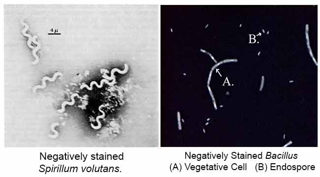
The main purpose of Negative staining is to study the morphological shape, size and arrangement of the bacteria cells that is difficult to stain. eg: Spirilla. It can also be used to stain cells that are too delicate to be heat-fixed.
It is also used to prepare biological samples for electron microscopy. It is used to view viruses, bacteria, bacterial flagella, biological membrane structures and proteins or protein aggregates, which all have a low electron-scattering power. It is also used for the study and identification of aqueous lipid aggregates like lamellar liposomes (le), inverted spherical micelles (M) and inverted hexagonal HII cylindrical (H) phases by Negative staining transmission electron microscopy.
Principle of Negative Staining
Negative staining requires an acidic dye such as India Ink or Nigrosin.
India Ink or Nigrosin is an acidic stain. This means that the stain readily gives up a hydrogen ion (proton) and the chromophore of the dye becomes negatively charged. Since the surface of most bacterial cells is negatively charged, the cell surface repels the stain. The glass of the slide will stain, but the bacterial cells will not. The bacteria will show up as clear spots against a dark background.
Reagents of Negative Staining
India ink
NigrosinNigrosin 100 gm/L, Formalin 5 ml/L in water
Procedure of Negative Staining
1. Place a very small drop (more than a loop full, less than a free falling drop from the dropper) of nigrosin near one end of a well-cleaned and flamed slide.
2. Remove a small amount of the culture from the slant with an inoculating loop and disperse it in the drop of stain without spreading the drop.
3. Use another clean slide to spread the drop of stain containing the organism using the following technique.
4. Rest one end of the clean slide on the center of the slide with the stain. Tilt the clean slide toward the drop forming an acute angle and draw that slide toward the drop until it touches the drop and causes it to spread along the edge of the spreader slide. Maintaining a small acute angle between the slides, push the spreader slide toward the clean end of the slide being stained dragging the drop behind the spreader slide and producing a broad, even, thin smear.
5. Allow the smear to dry without heating.
6. Focus a thin area under oil immersion and observe the unstained cells surrounded by the gray stain.
Procedure to view in Transmission Electron Microscope (TEM)
Hold a coated grid flim side up in a pair of self clamping forceps.
Make a 1:1 mixture of sample and negative stain (eg. 2% uranyl acetate or 2% sodium or potassium phosphotungstate, pH 7.4). Add 5µl to the grid. Smaller particles adsorb to the grid surface more rapidly than larger particles. Alternatively the sample mixed with fixative can be added to the grid before subsequent negative staining.
Incubate for 30-90 seconds then remove excess liquid with the torn edge of a piece of filter paper.
Air dry and examine in the TEM.
Results of Negative Staining
Negative Staining- Principle, Procedure and Result Interpretation
Objectives of Negative Staining
To perform a negative staining procedure.
To understand the benefit obtained from visualizing unstained microorganisms.
Principle of Negative Staining
Negative staining requires the use of an acidic stain such as India ink or nigrosin. The acidic stain, with its negatively charged chromogen, will not penetrate the cells because of the negative charge on the surface of bacteria. Therefore, the unstained cells are easily discernible against the colored background.
The practical application of negative staining is twofold.
First, since heat fixation is not require and the cells are not subjected to the distorting effects of chemicals and heat, their natural size and shape can be seen.
Second, it is possible to observe bacteria that are difficult to stain, such as some spirilla. Because heat fixation is not done during the staining process, keep in mind that the organisms are not killed and slides should be handled with care.
Reagent and Equipment’s for Negative Staining Nigrosin, Microincinerator or Bunsen burner, inoculating loop, staining tray, glass slides, lens paper, and microscope.
Procedure of Negative Staining
Place a small drop of nigrosin close to one end of a clean slide.
Using aseptic technique, place a loopful of inoculum from the bacterial culture in the drop of nigrosin and mix.
Place a slide against the drop of suspended organisms at a 45° angle and allow the drop to spread along the edge of the applied slide.
Push the slide away from the drop of suspended organisms to form a thin smear. Air-dry. Note: Do not heat fix the slide.
Negative Staining- Principle, Procedure and Result Interpretation
(Visited 1,046 times, 5 visits toda
Ex 6 - Negative Staining
What is the Negative Stain Used For?
Negative staining allows the morphology, arrangement and size of bacteria, to be viewed microscopically.
How Do Negative Stains Work?
Negative staining allows the morphology, arrangement and size of bacteria, to be viewed microscopically.
The negative stain uses the dye nigrosin, which is an acidic dye. Acids donate hydrogen ions, which are positively charge protons. As acids loose positive charge, the chromophores of the dye becomes negatively charged. The cell wall of the bacteria is naturally negatively charged. A rule of chemistry, is that opposite charges attract, and "like charges" (charges that are alike) repel one another. The result is that the bacteria cell remains unstained an appears as a clear object on the slide, while the area surrounding the bacteria cell is darkly stained by the nigrosin stain (also known as "Indian Ink").
How do you determine whether a stain is basic or acidic?
If the chromophore is positive, it is basic
If the chromophore is negative, it is acidic
What is considered a simple stain?
Staining procedures that use only one stain
List the steps for preparing a smear.
1-Place a loopful on water on the slide, 2-place a small amount of the culture onto the slide and mix, 3-allow smear to air dry, 4-flame the slide
List the steps of preparing a simple stain.
1-Cover smear with methylene blue for 30 seconds, 2-Wash with DI water
Of what value is a simple stain?
Determining cell morphology, size, and arrangement
What is the purpose of fixing the smear preparation?
Denatures bacterial enzymes
In heat fixing, what would happen if too much heat were applied?
It would damage the cell's structure and can even crack the slide.
Can dyes other than methylene blue be used for direct staining?
Yes any positively charged dye can be used such as safranin and basic fuchsin
Bacteria can be seen without staining, so why is fixing and staining important for microbiology?
It allows for later reexamination
What does the negative stain technique stain?
Background (the area around the cell, but NOT the cell itself)
Why doesn't a negative stain stain the bacteria?
Ionic repulsion; like charges repel. The bacteria cell wall and the chromophores of the acidic stain both have negative charges.
In which procedure are no heat fixing or strong chemicals used and why not?
Negative stain; to keep the bacteria from getting as distorted
When is the negative stain technique especially useful?
When other staining techniques don't clearly show cell morphology or size
List the steps of the negative staining technique.
1-Place a drop of nigrosin on slide, 2-Mix a loopful of broth culture with the nigrosin (no extra water is needed when the culture is taken from solid media as nigrosin contains a lot of water already), 3-Use a second slide to sweep the mixture across the first slide, making a color gradient, 4-Let the smear air dry
How does bacteria from a negative stain differ from that of a simple stain?
It's clearer because only the background is stained
Why is the size more accurate in a negative stain than in a simple stain?
The lack of heat fixing or strong chemicals keeps from distorting the cell
Could any dye be used in place of nigrosin in a negative stain?
No
What type of dye is used for negative staining? (Acidic or Basic)
Acidic
What kind of stain is the gram stain? How does is allow you to classify bacteria?
Differential; as either gram positive or gram negative
Describe the discovery of the gram stain technique.
Discovered accidentally by Hans Christian Gram in 1884 when he attempted to stain cells and found that some lost their color when the excess stain was washed off
In a gram stain, what are bacteria called when they are easily decolorized?
Gram negative
In a gram stain, what are bacteria called when they retain the primary stain?
Gram positive
Why do bacteria stain differently in a gram stain?
Chemical and physical differences in their cell walls
In a gram stain, what type of cell does this describe?
Crystal violet is picked up by the cell. Iodine reacts with the dye in the cytoplasm to form CV-I. The decolorizing agent dissolves the outer lipopolysaccharide layer, and the CV-I washes out through the thin layer of peptidoglycan.
Gram negative
When is the gram stain most consistent?
When done on cultures less than 24 hours old
List the steps of the gram stain technique.
1-Cover smear with crystal violet for 30 seconds and rinse, 2-Cover smear with Gram's iodine for 10 seconds and rinse, 3-Decolorize with alcohol and rinse, 4-Cover smear with safranin for 30 seconds and rinse
Name some gram positive bacteria.
S. aeru, S. epi, S. pyogenes, B. sub, B, meg
Name some gram negative bacteria.
E. coli, P. vulg, Neisseria
Why will old gram positive cells stain gram negative?
They can't retain the primary stain because with time the lipid layer degrades allowing the stain to reach the peptidoglycan
Can iodine be added before the primary stain in a Gram stain?
No
What color does Crystal Violet turn each of the cells in a gram stain?
Purple
What color does Gram's iodine turn each of the cells in a gram stain?
Purple
What color does ethyl alcohol turn each of the cells in a gram stain?
Gram positive-purple; Gram negative-clear
What color does safranin turn each of the cells in a gram stain?
Gram positive-purple; Gram negative-red
Which step can be omitted without affecting determination of the gram stain reaction?
Safranin
Suppose you gram stained a sample from a pure culture of bacteria and observed a field of red and purple cocci. Adjacent cells were not always the same color. What do you conclude?
The culture is old
Suppose you are viewing a gram stained field of red rods and purple cocci through the microscope. What do you conclude?
The culture has been cross-contaminated
Since you can't identify bacteria from a gram stain, why might a physician perform a gram stain on a sample before perscribing an antibiotic?
Because only certain antibiotics work depending on if the bacteria is gram positive or negative
What type of antibiotics work on gram positive bacteria?
Penicillin
What type of antibiotics work on gram negative bacteria?
Tetracycline
If you gram stained human cells, what would happen?
Primary stain would wash away because humans do not have cell walls
What type of stain is the acid-fast stain?
Differential
What is the value of the acid-fast stain?
Allows diagnosing of Mycobacterium and Nocardia, which do not decolorize with acid alcohol
The cell walls of acid-fast organisms have a wax-like lipid called mycolic acid, which does what?
Makes the cell wall impermeable to most stains
What the difference in the Ziehl-Neelsen acid-fast stain and the Kinyoun acid-fast stain?
In the Kinyoun stain (cold stain), the concentrations of phenol and carbolfuchsin are increased so heating isn't necessary
List the steps of the acid-fast stain.
1-Cover smear with carbolfuchsin for 5 minutes and rinse, 2-Wash smear with acid-alcohol on and off and rinse, Cover smear with Brilliant Green K (Methylene Blue) for 1 minute and rinse
What color will an acid-fast positive bacteria be?
Red
What color will an acid-fast negative bacteria be?
Green
Name an acid-fast negative bacteria
E. coli
Name an acid-fast positive bacteria
M. phlei
What is the decolorizing agent in the Gram stain? In the acid-fast stain?
Ethyl alcohol; Acid-alcohol
What diseases are diagnosed using the acid-fast staining technique?
Tuberculosis and Leprosy
What is phenol and what percent is its usual application?
Disinfectant; 5%
How might the acid-fast characteristic of Mycobacterium enhance the organism's ability to cause disease?
The lipids in their cell wall give extra protection; they are not easily phagocytized
Clinical specimens suspected of containing Mycobacterium are digested with sodium hydroxide for 30 minutes prior to staining. Why is this technique used? Why isn't this technique used for staining other bacteria?
To remove unwanted bacteria from specimen; the sodium hydroxide would kill other bacteria
What are the most familiar genera that form endospores?
Bacillus and Clostridium
Why are endospores called resting bodies?
Because they do not metabolize and are resistant to many harsh conditions
When are endospores formed?
When essential nutrients or water are not available
What happens to the cell when an endospore is formed?
It disintegrates
Many bacteria secrete chemicals that adhere to their surfaces, forming a viscous coat. What is this structure called when it is round or oval shaped? Irregularly shaped and loosely bound?
Capsule; Slime layer
How is a bacteria's ability to form a capsule determined?
Genetically
How is the size of a bacteria's capsule determined?
The medium it's growing on
What are most capsules composed of?
Polysaccharides, which are uncharged
Because of a capsules nonionic nature, _____ stains will not adhere to it.
Simple
How must a capsule be stained in order to view it under a microscope?
The background must be stained, leaving the capsules unstained
Capsules have a very important role in the _____ of some bacteria.
Virulence
How do capsules contribute to the virulence of some bacteria?
Capsules are not easily phagocytized
These thin proteinaceous structures that originate in the cytoplasm and project out from the cell wall are the most common means of motility in bacteria
Flagella
List the steps of an endospore stain.
1-Place a piece of paper towel over the slide, 2-Cover the paper with malachite green, 3-Steam the slide for 5 minutes then rinse, 4-Cover smear with safranin for 30 seconds and rinse
List the steps of a capsule stain.
1-Place a loopful of congo red on slide, 2-Put a thick smear of bacteria in the congo red and mix, 3-Let smear air dry, 4-Drip acid-alcohol on slide for 15 seconds and rinse, 5-Cover smear with acid fuchsin for 1 minute and rinse
What are the gram reactions of Clostridium and Bacillus?
Both are gram positive
How might flagella contribute to pathogenicity?
They help evade the immune response
In an endospore stain, how would slides of 24-hour old Bacillus and 72-hour old Bacillus differ?
At 72 hours, endospores would have formed
Of what morphology are most bacteria possessing flagella?
Bacillus
Which morphology does not usually have flagella?
Coccus
What prevents the cell from appearing green in the finished endospore stain?
Vegetative cells are easily decolorized so the green dye washes off
You can see endospores by simple staining. Why not use this technique?
It would be more difficult to pick them out because they would be transparent
How would an endospore stain of Mycobacterium appear?
Mycobacterium's waxy cell wall would retain the green dye, suggesting that they have endospores when they actually do not
What type of culture medium would increase the size of a bacterial capsule?
A medium rich in polysaccharides
Describe the microscopic appearance of encapsulated Streptococcus if stained with safranin and nigrosin.
The bacteria would be red, the capsule clear, and the background purplish-black
In the Dorner endospore stain, a smear covered with carbolfuchsin is steamed, then decolorized with acid-alcohol and counterstained with nigrosin. Describe the microscopic appearance after this procedure.
The endospores should be very bright pink, the bacteria light pink, and the background purplish-black
What makes an agar deep useful?
Some oxygen prefer to grow in less oxygen
What are two ways to determine motility of a bacteria?
Using an agar deep or the hanging drop technique
What evolutionary advantage would there be to the formation of a pellicle in a liquid medium by a bacterium?
They could form colonies and have a greater chance of survival
How can you tell that a medium is sterile?
Compare it with a control
Why is it desirable that microscope lenses be parfocal?
So you don't have to refocus everytime you change objectives
What controls the amount of light reaching the ocular lens?
Diaphragm
Is you lens corrected for chromatic aberrations?
Yes
Name two ways in which you can enhance the resolving power of a microscope.
Decrease the wavelength of light and increase the numerical aperture
What would occur if water were accidentally used in place of immersion oil?
It would not be as clear because water does not have the same refractive index as glass
What advantages does the low power objective have over the oil immersion objective for viewing fungi?
Fungi cells are large so you can see more of the cell
Organisms Used:
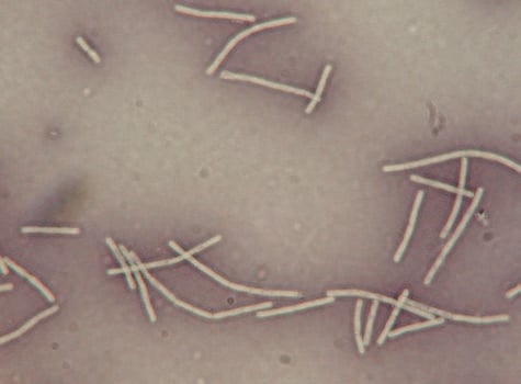
Escherichia coli (E. coli) - Gram (-) rod
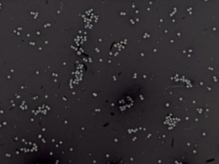
Staphylococcus aureus (Staph. aureus)
Staphylococcus aureus is a gram-positive bacteria that is roundGram (+) cocci
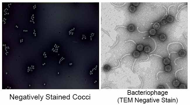
Negative staining is a simple and rapid method for studying the morphology and ultrastructure of small particulate specimens. Samples stained in this manner show great structural integrity because the stains used not only delineate the ultrastructure, but also act as a fixative, thus protecting samples from irradiation damage by the electron beam. Negative staining also reduces the surface-tension forces at the air-liquid interface, thus reducing shrinkage and specimen collapse. These effects are promoted by the heavy metal salts used in negative stains. While negative staining is one of the oldest techniques for studying the ultrastructure of particulate samples at the electron microscopy level, it has never been surpassed by any other technique for determining the surface structures, size, and shapes of specimens such as viruses, bacteria, and macromolecules, with resolution down to the 2-nm level.
Nevertheless negative staining can be used to visualize bacteriophages and their interaction with the membrane of proteoliposomes containing their receptor. Also important is that full capsids of bacteriophages that contain DNA can be distinguished readily from empty capsids. Indeed, filled capsids have a characteristic icosahedral shape, whereas empty heads appear broken and retain a lower amount of stain. Thus, it is possible to visualize the content of the capsid heads and determine the percentage of the heads that are full, empty, or partially empty. For example, it has been shown, in the case of λ phage, that 75% of phages bound to their receptor appear to have ejected all or part of their DNA at 37°, whereas at 4° most of the phages are still bound to the liposome, with their heads full of DNA.
NEGATIVE STAINING – INTRODUCTION, PRINCIPLE, PROCEDURE,
RESULTS & APPLICATIONS
Posted By: SAHIL BATRA
APPLICATIONS OF NEGATIVE STAINING, FACTS OF NEGATIVE STAINING, NEGATIVE STAIN, NEGATIVE STAINING, NEGATIVE STAINING METHOD, NEGATIVE STAINING PROTOCOL,
NEGATIVE STAINING TECHNIQUE, PRINCIPLE OF NEGATIVE STAINING, PROCEDURE OF NEGATIVE STAINING, RESULTS OF NEGATIVE STAINING, USES OF NEGATIVE STAINING, WHAT
IS NEGATIVE STAIN, WHAT IS THE PURPOSE OF NEGATIVE STAINING, WHY DO WE DO NEGATIVE STAINING
INTRODUCTION TO NEGATIVE STAINING TECHNIQUE
Sometimes it is more convenient to determine the overall bacterial cell morphology without any use of harsh
staining or heat fixing techniques that changes the shape of the bacterial cells.
The negative staining is preferred in cases when the bacterium does not stain well (e.g. some of the spirochetes)
or when it is desirable to confirm observations made on the shape and size or Morphology of the bacteria
observed either in a wet mount or hanging drop preparations etc.
Negative staining is also good for viewing capsules but preferably Capsule staining is done in that cases.
MICROBIOLOGY PRACTICALS MICROBIOLOGY STAININGS
/
81
10/10/2018 Negative Staining Technique - Principle, Proc
10/10/2018 Negative Staining Technique - Principle, Procedure, Results | Microbiology Practicals
https://paramedicsworld.com/microbiology-stainings/negative-staining-technique-principle-procedure-results/medical-paramedical-studynotes#.W72q… 3/11
⇒ Mix well the culture with dye taken on the Glass slide.
⇒ Now, take another Microscopic Glass slide, place it near to the specimen-dye mixture at an angle of about 30°
– 45°.
NEGATIVE STAINING SPREADER METHOD
STEP 2
⇒ Move the slide toward the drop of the specimen-dye mixture until the contact is made with the drop at the
specific angle. Then move the spreader slide smoothly and rapidly forward over the specimen slide, drawing the
dye mixture behind it into a thin film.
NEGATIVE STAINING SPREADER METHOD
STEP 3
Alternatively,
⇒ Take a drop of nigrosine or India ink and place it in the middle of the Clean & Grease free Glass slide.
NEGATIVE STAINING LOOP METHOD STEP 1
⇒ Now, with the help of Sterilized inoculating loop transfer a small portion of the specimen to the slide
containing a Drop of Dye.
Lab Microscope Study Notes
Laboratory Test Cell Culture
10/10/2018 Negative Staining Technique - Principle, Procedure, Results | Microbiology Practicals
https://paramedicsworld.com/microbiology-stainings/negative-staining-technique-principle-procedure-results/medical-paramedical-studynotes#.W72q… 4/11
NEGATIVE STAINING LOOP METHOD STEP 2
⇒ Mix well the Specimen with the Dye using the sterilized straight wire and spread evenly over an area of about
1 – 2 cm.
NEGATIVE STAINING LOOP METHOD STEP 3
⇒ Allow the smear to Air dry and then observe under the microscope at High power objective (45X) and oil
immersion (100X) objectives.
NEGATIVE STAINING LOOP METHOD STEP 4
OBSERVATIONS & RESULTS ON NEGATIVE STAINING
In Negative staining Preparations, the Bacterial cells observed as the Clear transparent bodies or objects, may
be of variable size and shape if you are using a mixture of bacteria, against a dark background.
VEGETATIVE CELLS & SPORES OF BACILLI ARE CLEARLY VISIBLE IN NEGATIVE STAIN PREPARATION
FACTS ABOUT NEGATIVE STAINING
10/10/2018 Negative Staining Technique - Principle, Procedure, Results | Microbiology Practicals
https://paramedicsworld.com/microbiology-stainings/negative-staining-technique-principle-procedure-results/medical-paramedical-studynotes#.W72q… 5/11
⇒ In Negative staining method, we stain the background and not the bacterial cells so after drying there is no
need of washing step and the slide is directly observed under oil immersion objectives (100 X).
⇒ In Negative staining method, heat fixation step is avoided because heat fixation can alter morphological
characters of the bacterial cell, melt the capsule or slime layer and distorts the actual morphology.
NEGATIVE STAIN PREPARATION SHOWING COCCI IN CLUSTERS
APPLICATION OF NEGATIVE STAINING
⇒ This method is commonly used to study the morphological characters of cell that includes the size, shape, and
arrangement of bacterial cells without distorting their actual characters.
⇒ This method is quite easy and useful in observing the bacterial cells that are difficult to stain by other staining
techniques. For e.g. – Spirillum, Spirochetes etc.
⇒ This Staining method can also be used to observe the capsules of the bacteria.
That’s all about the Negative Staining Technique – Introduction, Principle, Procedure, Results
& Application


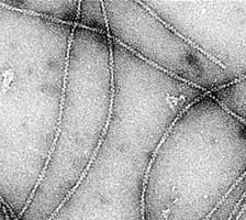 Negatively stained actin filaments. Photo: Craig Lab.
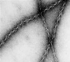 Negatively stained actomyosin filaments.Photo: Craig Lab.