Bacterial Spores: Structure, Importance and examples of spore forming bacteria

Bacterial spores are highly resistant, dormant structures (i.e. no metabolic activity) formed in response to adverse environmental conditions. They help in the survival of the organisms during adverse environmental conditions; they do not have a role in reproduction.
Note: Spores of fungi have a reproductive role.
Spore formation (sporulation) occurs when nutrients, such as sources of carbon and nitrogen are depleted. Bacterial spores are highly resistant to
- Heat
- Dehydration
- Radiation and
- Chemicals.
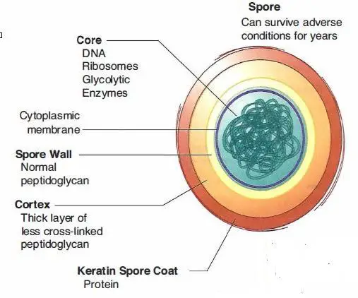
An endospore is structurally and chemically more complex than the vegetative cell. It contains more layers than vegetative cells. Resistance of Bacterial spore may be mediated by dipicolinic acid, a calcium ion chelator found only in spores. When the favorable condition prevail, (i.e. availability of water, appropriate nutrients) spores germination occurs which forms vegetative cells of pathogenic bacteria.
Following factors/constituents plays major role for the resistance of Bacterial Spore:
- Calcium dipicolinate in core
- Keratin spore coat
- New enzymes (i.e., dipicolinic acid synthetase, heat-resistant catalase)
- Increases or decreases in other enzymes.
A mature endospore contains a complete set of the genetic material (DNA) from the vegetative cell, ribosomes and specialized enzymes.
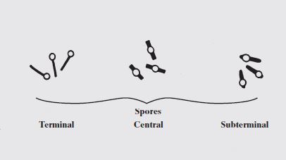
The shape and the position of spores vary in different species and can be useful for classification and identification purposes. Endospores may be located in the middle of the bacterium (central), at the end of the bacterium (terminal) and near the end of the bacteria (subterminal) and may be spherical or elliptical.
Spores may be:
- Central or equatorial, giving the bacillus aspindle shape (eg. Clostridium bifermentans)
- Sub-terminal, the bacillus appearing Club shaped (eg. Clostridium perfringens)
- Oval and terminal, resembling a tennis racket (eg. Clostridium tertium)
- Spherical and terminal, giving a drumstick appearance (Clostridium tetani)
Mature endospores are released from the vegetative cell to become free endospores. When the free endospores are placed in an environment that supports growth, the endospores will revert back to a vegetative cell in a process called germination. It should be noted that unlike the process of binary fission observed with vegetative cells, endospore formation is not a reproductive process but a process of differentiation that provides the bacteria with a mechanism for survival.
Constituents of Bacterial Spores:
- Thick keratinlike coat
- Peptidoglycan
- Cell membrane
- A small amount of cytoplasm
- Very little water
- Bacterial DNA
Medical Importance of Bacterial Spores
| Important features of Spores | Medical Implications |
| Spores are highly resistant to heating; spores are not killed by boiling (100OC) but are killed at 121OC. | Medical supplies must be heated to 121oC for atleast 15 minutes to be sterilized. |
| Spores are highly resistant to many chemicals, including most disinfectants. | Only solution designated as sporicidal will kill spores. |
| Spores can survive for many years in soil and other inanimate objects. | Wound contaminated with soils can be infected with spores and cause diseases such as tetanus, gas gangrene. |
| Spores do not exhibit measurable metabolic activity. | Antibiotics are ineffective against spores. |
| Spores formed only when nutrients are insufficient. | Spores are not often found at the site of infection because nutrients are not limiting. |
Examples of Spore forming Bacteria- Spores formed by only two genera of Gram positive rods are of medical importance.
- Bacillus spp
- Clostridium spp
Endospore Staining: Principle, Procedure and Results

When vegetative cells of certain bacteria such as Bacillus spp and Clostridium spp are subjected to environmental stresses such as nutrient deprivation, they produce metabolically inactive or dormant form-endospore. Formation of endospore circumvent the problems associated with environmental stress and helps them to survive.
During unfavorable conditions (especially when carbon and nitrogen become unavailable) endospores can form within different areas of the vegetative cell. They can be central, subterminal, or terminal. Central endospores are located within the middle of the vegetative cell. Terminal endospores are located at the end of the vegetative cell. Sub-terminal endospores are located between the middle and the end of the cell.
Most endospore forming bacteria are found in soil or aquatic environments. However, some species of Bacillus and Clostridium have medical significance. Clostridium perfringens, C. botulinum (a potential agent of bioterrorism) and C. tetani are the causative agents of gas gangrene, botulism and tetanus, respectively. Bacillus anthracis and Bacillus cereus are the causative agents of anthrax and a self limiting food poisoning, respectively.
Principle of Spore Staining:
A differential staining technique (the Schaeffer-Fulton method) is used to distinguish between the vegetative cells and the endospores. A primary stain (malachite green) is used to stain the endospores. Because endospores resist staining, the malachite green will be forced into (i.e, malachite green permeate the spore wall) the endospores by heating. In this technique heating acts as a mordant.
There is no need of using any decolorizer in this spore staining as the primary dye malachite green bind relatively weakly to the cell wall and spore wall .In fact If washed well with water the dye come right out of cell wall however not from spore wall once the dye is locked in. Water is used to decolorize the vegetative cells.
( Note: In Gram Staining and AFB Staining we use Alcohol or Acid Acohol or Acid as a decolorizer but in spore staining water is sufficient ( to be used as decolorizer) because:
- malachite green dye is water-soluble and does not adhere well to the cell wall
- vegetative cells have been disrupted by heat,
because of these reasons, the malachite green rinses easily from the vegetative cells. )
As the endospores are resistant to staining, the endospores are equally resistant to de-staining and will retain the primary dye while the vegetative cells will lose the stain. The addition of a counterstain or secondary stain (safranin) is used to stain the decolorized vegetative cells.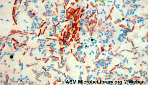
When visualized under microscopy the cells should have three characteristics:
- the vegetative cells should appear pink/red (i.e. color of counter stain),
- the vegetative cells that contain endospores should stain pink while the spores should be seen as green ellipses within the cells.
- Mature, free endospores should not be associated with the vegetative bacteria and should be seen as green ellipses.
Procedure of endospore stain:
- Prepare smears of organisms to be tested for presence of endospores on a clean microscope slide and air dry it.
- Heat fix the smear.
- Place a small piece of blotting paper (absorbent paper) over the smear and place the slide (smear side up) on a wire gauze on a ring stand.
- Heat the slide gently till it starts to evaporate (either by putting the slide on a staining rack that has been placed over a boiling water bath or via bunsen burner).
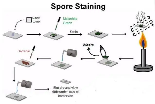
Spore staining procedure - Remove the heat and reheat the slide as needed to keep the slide steaming for about 3-5 minutes. As the paper begins to dry add a drop or two of malachite green to keep it moist, but don’t add so much at one time that the temperature is appreciably reduced.
# DO NOT OVERHEAT. The process is steaming and not baking. - After 5 minutes carefully remove the slide from the rack using a clothespin
- Remove the blotting paper and allow the slide to cool to room temperature for 2 minutes.
- Rinse the slide thoroughly with tap water (to wash the malachite green from both sides of the microscope slide).
- Stain the smear with safranin for 2 minutes.
- Rinse both side of the slide to remove the secondary stain and blot the slide/ air dry.
Observe the bacteria under 1000X (oil immersion) total magnification.
Results: The vegetative cells will appear pink/red and the spores will appear green.